Home
/ Diagram Cross Section Of A Bone : Medical Diagram Of The Structure Of The Inside Cross Section Of The Tooth Stock Vector Illustration Of Medical Education 125924960 : For example, to read this diagram literally, since the cartilage can be seen inside the.
Diagram Cross Section Of A Bone : Medical Diagram Of The Structure Of The Inside Cross Section Of The Tooth Stock Vector Illustration Of Medical Education 125924960 : For example, to read this diagram literally, since the cartilage can be seen inside the.
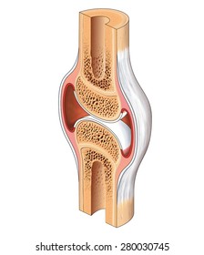
Diagram Cross Section Of A Bone : Medical Diagram Of The Structure Of The Inside Cross Section Of The Tooth Stock Vector Illustration Of Medical Education 125924960 : For example, to read this diagram literally, since the cartilage can be seen inside the.. Diagram of a cross section of the coiled cochlea. Explaned distal and proximal epiphysis. This article covers the anatomy of the spinal cord including its structure tracts and function. Cross section of bone diagram. Diagram with articular cartilage, marrow, spongy bone, medullary cavity, endosteum, diaphysis, and periosteum. can be used for personal and commercial purposes.
Cochlea diagram cross section as the travellers or messenger terminals are normally interconnected, the prevalent terminal is the only a single left. For example, to read this diagram literally, since the cartilage can be seen inside the cutaway section of bone, it incorrectly indicates that the cartilage in fact goes through the bone structure, rather than just being found around the bone end. Diagram with articular cartilage, marrow, spongy bone, medullary cavity, endosteum, diaphysis, and periosteum. can be used for personal and commercial purposes. Cross section diagrams are used a lot by architects and engineers to show what a building or machine might look like before it's built. In the last decade, considerable technological improvements have been made to repair damaged bones and tissue, such as bone cross sections with implants for microscopic examinations.
Bone Cross Section High Res Stock Images Shutterstock from image.shutterstock.com Both types of bone marrow are enriched with blood vessels and capillaries. Bone cross section diagram ipad folio cases. Diagram with articular cartilage, marrow, spongy bone, medullary cavity, endosteum, diaphysis, and periosteum. can be used for personal and commercial purposes. A cross section of a human long bone. This article covers the anatomy of the spinal cord including its structure tracts and function. Diagram with articular cartilage, marrow, spongy bone, medullary cavity, endosteum, diaphysis, and periosteum. They build the entire picture, improve your understanding, consolidate the information and facilitate recall. Spongy bone and compact bone.
(micrograph provided by the regents of university of michigan.
They build the entire picture, improve your understanding, consolidate the information and facilitate recall. There are trabeculae in spongy bone which gives its sponge like appearance. Cross section of bone diagram. The surface features of bones vary considerably, depending on the function and location in the body. Volume of a solid figure with uniform cross section. From wikimedia commons, the free media repository. Diagram with articular cartilage, marrow, spongy bone, medullary cavity, endosteum, diaphysis, and periosteum. can be used for personal and commercial purposes. This article covers the anatomy of the spinal cord including its structure tracts and function. Ga voor hoogwaardige illustratieve kunst met een hoge resolutie naar getty images. Metaphseal region on the left, diaphyseal region on the right. Cross section diagrams are used a lot by architects and engineers to show what a building or machine might look like before it's built. Blood vessels and nerves enter the bone through the. (micrograph provided by the regents of university of michigan.
Metaphseal region on the left, diaphyseal region on the right. The surface features of bones vary considerably, depending on the function and location in the body. For example, to read this diagram literally, since the cartilage can be seen inside the. Cross section diagrams a cross section diagram is if you would take a knife and cut through one side of a diagram to see the inside and outside in one picture. Whereas a long bone has only one layer of compact bone (see fig 1).
What Is The Structure And Function Of The Compact Bone Socratic from cronodon.com Both types of bone marrow are enriched with blood vessels and capillaries. Blood vessels and nerves enter the bone through the. Cross section of a human bone. In a cross section of a bone we can see two types of bone tissue: Volume of a solid figure with uniform cross section. The surface features of bones vary considerably, depending on the function and location in the body. Compact bone is the outer layer and the spongy bone forms the inner layer. In the last decade, considerable technological improvements have been made to repair damaged bones and tissue, such as bone cross sections with implants for microscopic examinations.
Jump to navigation jump to search.
Two types of bone tissues in cross section of a long bone : Cross section of a bone diagram : They build the entire picture, improve your understanding, consolidate the information and facilitate recall. Bekijk hoogwaardige illustraties van crosssection diagram of a human long bone. Spongy bone and compact bone. There are trabeculae in spongy bone which gives its sponge like appearance. Function of bone bone is a living, metabolically active and highly organized tissue consisting of a. Diagram of a cross section of the coiled cochlea. Jump to navigation jump to search. Explaned distal and proximal epiphysis. In a cross section of a bone we can see two types of bone tissue: Bone is found in the shafts of long bone and consists of various cylindrical units named as haversian system 47. Diagram with articular cartilage, marrow, spongy bone, medullary cavity, endosteum, diaphysis, and periosteum.
Diagram with articular cartilage, marrow, spongy bone, medullary cavity, endosteum, diaphysis, and periosteum. Diagram with articular cartilage, marrow, spongy bone, medullary cavity, endosteum, diaphysis, and periosteum. Diagram of blood and nerve supply to bone. As the names suggest compact bone looks compact and the spongy bone looks like skull bone is a flat bone. For example, to read this diagram literally, since the cartilage can be seen inside the cutaway section of bone, it incorrectly indicates that the cartilage in fact goes through the bone structure, rather than just being found around the bone end.
Bone Structure Anatomy And Physiology I from s3-us-west-2.amazonaws.com Whereas a long bone has only one layer of compact bone (see fig 1). Volume of a solid figure with uniform cross section. Hope you enjoy and please. Diagram with articular cartilage, marrow, spongy bone, medullary cavity, endosteum, diaphysis, and periosteum. Both types of bone marrow are enriched with blood vessels and capillaries. Explaned distal and proximal epiphysis. Ear external and internal anatomy cross section unlabeled stock illustration 9895a hr fotosearch / wh. The surface features of bones vary considerably, depending on the function and location in the body.
Function of bone bone is a living, metabolically active and highly organized tissue consisting of a.
Both types of bone marrow are enriched with blood vessels and capillaries. Bekijk hoogwaardige illustraties van crosssection diagram of a human long bone. Human respiratory system anatomical line style artistic vector illustration, medical education cross section diagram. Diagram with articular cartilage, marrow, spongy bone, medullary cavity, endosteum, diaphysis, and periosteum. As the names suggest compact bone looks compact and the spongy bone looks like skull bone is a flat bone. Diagram of a cross section of the coiled cochlea. Patient's and clinic's names removed real brain mri slide of a young woman. Jump to navigation jump to search. In the last decade, considerable technological improvements have been made to repair damaged bones and tissue, such as bone cross sections with implants for microscopic examinations. Cross section of bone diagram. As shown in figure 2. For example, to read this diagram literally, since the cartilage can be seen inside the cutaway section of bone, it incorrectly indicates that the cartilage in fact goes through the bone structure, rather than just being found around the bone end. In a cross section of a bone we can see two types of bone tissue:
The cross section of a solid is a plane section resulting from a cut (real or imaginary) perpendicular to the length (or breadth of height) of the solid cross section of a bone. Find the perfect bone diagram stock illustrations from getty images.

